
 Researchers from the U.S. National Institutes of Health have collaborated with NVIDIA experts on an AI-accelerated method to monitor COVID-19 disease severity over time from patient chest CT scans.
Researchers from the U.S. National Institutes of Health have collaborated with NVIDIA experts on an AI-accelerated method to monitor COVID-19 disease severity over time from patient chest CT scans.
Published today in Scientific Reports, this work studied the progression of lung opacities in chest CT images of COVID patients, and extracted insights about the temporal relationships between CT features and lab measurements.
Quantifying CT opacities can tell doctors how severe a patient’s condition is. A better understanding of the progression of lung opacities in COVID patients could help inform clinical decisions in patients with pneumonia, and yield insights during clinical trials for therapies to treat the virus.
Selecting a dataset of more than 100 sequential chest CTs from 29 COVID patients from China and Italy, the researchers used an NVIDIA Clara AI segmentation model to automate the time-consuming task of segmenting the total lung in each CT scan. Expert radiologists reviewed the total lung segmentations, and manually segmented the lung opacities.
To track disease progression, the researchers used generalized temporal curves, which correlated the CT imaging data with lab measurements such as white blood cell count and procalcitonin levels. They then used 3D visualizations to reconstruct the evolution of COVID opacities in one of the patients.
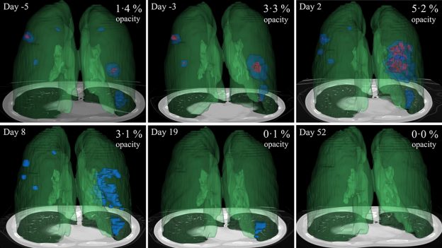
The team found that lung opacities appeared between one and five days before symptom onset, and peaked a day after symptoms began. They also analyzed two opacity subtypes — ground glass opacity and consolidation — and discovered that ground glass opacities appeared earlier in the disease, and persisted for a time after the resolution of the consolidation.
In the paper, the researchers showed how CT dynamic curves could be used as a clinical reference tool for mild COVID-19 cases, and might help spot cases that grow more severe over time. These curves could also assist clinicians in identifying chronic lung effects by flagging cases where patients have residual opacities visible in CT scans long after other symptoms dissipate.
This paper follows research published in Nature Communications, in which the team used deep learning to distinguish COVID-19 associated pneumonia from non-COVID pneumonia in chest scans. The deep learning models were developed using the NVIDIA Clara application framework for medical imaging, and are available for research use in the NGC catalog.
Read the full paper in Scientific Reports. Download the models from NGC and visit our COVID-19 research hub for more.
Learn more about NVIDIA’s work in healthcare at the GPU Technology Conference, April 12-16. Registration is free. The healthcare track includes 16 live webinars, 18 special events, and over 100 recorded sessions.
Subscribe to NVIDIA healthcare news.